

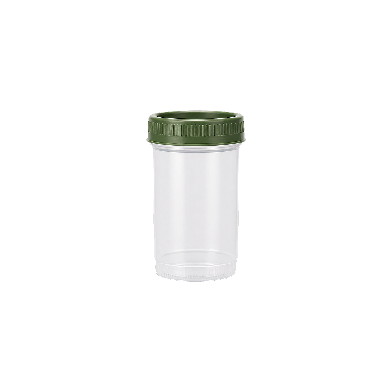
Urinalysis, Complete Test with Reflex to Urine Culture
A broad screen of the urine that flags infection, kidney issues, blood sugar problems, and dehydration in one sample.
Consider this test if:
- Burning, frequent, or urgent urination, or pelvic and flank pain that could signal a UTI
- Visible blood in the urine, foamy urine, or unexplained swelling
- Diabetes or blood-sugar concerns, checking for glucose or ketones in the urine
- Establishing a kidney and urinary baseline as part of a general checkup
- Tracking known kidney issues or recurrent infections over time
- HSA/FSA eligible
- Results delivered to your dashboard · Reviewed by a real clinician
- No blood draw needed. Urine sample processed by a CLIA/CAP-accredited lab · Find labs
Reflex testing
If your urinalysis shows signs of a possible infection, a urine culture runs automatically to identify the organism and guide treatment. This follow-up is done at no additional cost.
Pre-test considerations
No fasting required. A clean-catch midstream sample gives the most reliable result. Avoid testing during menstruation, since blood can contaminate the sample and skew the microscopic findings. Very high vitamin C intake and some medications can affect certain dipstick readings.

Video consult with your Care Team
A 1:1 call to discuss your health.
What members say on Trustpilot
What this test is for
A complete urinalysis examines urine three ways: physical (color, clarity, concentration), chemical (dipstick for glucose, protein, ketones, blood, bilirubin, nitrite, leukocyte esterase, pH), and microscopic (red and white cells, bacteria, yeast, casts, crystals, and epithelial cells in the sediment). Together these point to urinary tract infections, kidney disease, poorly controlled blood sugar, dehydration, and liver or blood disorders. Protein or blood in the urine can be an early sign of kidney damage, glucose and ketones can reflect diabetes, and nitrites with white cells and bacteria suggest infection. It is commonly ordered to investigate painful or frequent urination, flank or back pain, visible blood, or as a general health and kidney baseline. Results are read alongside symptoms, since minor findings like a few crystals or trace protein are often benign.
Biomarkers tested
Includes 26 biomarkers
- Specimen
- Urine sediment
- Method
- Light microscopy
- Measures
- Presence / threshold
This test counts bacteria per high power field in urine sediment examined under a microscope, a direct look for organisms that shouldn't be living in your urinary tract. Elevated counts point to a urinary tract infection, especially alongside white blood cells, and often explain burning with urination, urgency, frequency, pelvic pain, or cloudy, foul-smelling urine. It's a quick way to confirm or rule out infection when symptoms flare, or to check that a prior UTI has actually cleared.
- Specimen
- Urine sediment
- Method
- Light microscopy
- Measures
- Count per area
This dipstick check flags whether bilirubin, a breakdown product of old red blood cells that your liver normally processes into bile, is leaking into your urine. Healthy urine carries none of it, so a positive result points to conjugated bilirubin backing up into the bloodstream, a sign of bile duct obstruction, hepatitis, or other liver dysfunction. It's a useful piece of the puzzle when you're investigating dark urine, yellowing skin or eyes, pale stools, itching, or unexplained fatigue, and it often shows up on the same strip as urobilinogen for a fuller read on liver and bile flow.
- Specimen
- Urine
- Method
- Test strip
- Measures
- Presence / threshold
This marker counts calcium oxalate crystals seen directly under the microscope in a spun sample of your urine, the same crystal type that forms most kidney stones. Finding them is common and often harmless, but frequent or heavy amounts point to concentrated urine, low fluid intake, or a diet high in oxalate (spinach, nuts, chocolate, tea), and correlate with stone risk over time. It's a useful check if you've had a kidney stone before, get recurrent flank pain or blood in your urine, or want to confirm that hydration and dietary changes are actually reducing your crystal load.
- Specimen
- Urine sediment
- Method
- Light microscopy
- Measures
- Count per area
- Specimen
- Urine sediment
- Method
- Light microscopy
- Measures
- Count per area
This test looks under the microscope at urine sediment to count crystals per high power field, the mineral and salt deposits that form when urine holds more of a substance than it can keep dissolved. Certain crystal types point to specific problems: calcium oxalate and uric acid crystals often accompany kidney stone formation, while others can flag dehydration, diet extremes, or liver and metabolic conditions. It's a useful add-on if you've had flank pain, blood in your urine, or a history of kidney stones, and useful for tracking whether hydration or dietary changes are actually reducing stone-forming risk.
- Specimen
- Urine sediment
- Method
- Light microscopy
- Measures
- Count per area
This dipstick test checks whether glucose is spilling into your urine, which normally shouldn't happen because your kidneys reclaim virtually all of it back into the blood. Glucose shows up in urine when blood sugar climbs high enough to overwhelm that reabsorption, most often from undiagnosed or poorly controlled diabetes. It's a quick, low-cost flag rather than a precise reading, so a positive result points you toward blood-based tests like fasting glucose or A1c to confirm what's actually happening.
- Specimen
- Urine
- Method
- Test strip
- Measures
- Presence / threshold
Granular casts are cylindrical clumps of degenerated cellular material that form in the kidney's tubules and wash out into urine, visible under the microscope during a urine sediment exam. A few granular casts can turn up after hard exercise or dehydration, but larger numbers point to tubular cell injury and often show up in acute kidney injury, dehydration, or chronic kidney disease. This test is useful for investigating unexplained changes in urine output, swelling, rising creatinine, or fatigue, and for tracking kidney recovery after an insult like illness, medication exposure, or a low-flow event.
- Specimen
- Urine sediment
- Method
- Light microscopy
- Measures
- Count per area
This dipstick screen detects hemoglobin in urine, the protein that carries oxygen in red blood cells and, when present in urine, signals that blood is leaking somewhere along the kidneys or urinary tract. A positive result points to causes ranging from kidney irritation, UTIs, kidney stones, or strenuous exercise, to more serious conditions like kidney disease that warrant follow-up. It is a simple way to catch silent urinary tract or kidney issues before symptoms like pain, changes in urination, or visible blood appear.
- Specimen
- Urine
- Method
- Test strip
- Measures
- Presence / threshold
Hyaline casts are protein molds that form in the kidney's tubules and wash into urine, and technicians count them under the microscope during a urine sediment exam. A few are normal, especially after exercise, dehydration, or a fever, so an isolated finding usually needs no action. A high number, particularly alongside protein in urine, reduced output, or symptoms like swelling and fatigue, points toward more concentrated urine or early kidney stress worth tracking with follow-up testing.
- Specimen
- Urine sediment
- Method
- Light microscopy
- Measures
- Count per area
This dipstick test detects ketones spilling into urine when the body burns fat for fuel instead of glucose. It's useful for tracking a ketogenic or low-carb diet, confirming your body has shifted into fat-burning mode, or checking on a fasting protocol. Unexplained positive results paired with high blood sugar, excessive thirst, or fatigue warrant prompt follow-up, since that combination can signal diabetic ketoacidosis rather than diet-driven ketosis.
- Specimen
- Urine
- Method
- Test strip
- Measures
- Presence / threshold
Leukocyte esterase is an enzyme released by white blood cells, and this dipstick test detects it in urine as a marker of inflammation in the urinary tract. A positive result signals that neutrophils have shown up to fight something, usually a urinary tract infection, and often shows up alongside symptoms like burning with urination, urgency, frequency, or pelvic discomfort. It pairs with urine nitrites and a urinalysis to confirm infection and decide whether further culture or treatment is needed.
- Specimen
- Urine
- Method
- Test strip
- Measures
- Presence / threshold
This dipstick reading detects nitrite in urine, produced when certain bacteria (E. coli and other common culprits) convert dietary nitrate into nitrite as they multiply. A positive result signals a likely urinary tract infection and pairs with leukocyte esterase to confirm it, useful when you're tracking down burning urination, urgency, frequency, or pelvic discomfort. It's also a quick proactive check if you're prone to recurrent UTIs and want to catch one early, before it climbs to the kidneys.
- Specimen
- Urine
- Method
- Test strip
- Measures
- Presence / threshold
This measures how acidic or alkaline your urine is, a quick readout of how your kidneys manage acid-base balance and a marker that shifts with diet, hydration, and metabolic state. Persistently low (acidic) urine is linked to kidney stone formation, particularly uric acid stones, while high (alkaline) urine points to certain infections or renal tubular issues and can favor calcium phosphate stones. If you've had kidney stones before, get recurring UTIs, or are dialing in a diet (high protein, keto, or heavy produce intake) and want to see how it's shifting your body's chemistry, this is a fast way to check.
- Specimen
- Urine
- Method
- Test strip
- Measures
- Concentration
This test counts red blood cells per high-power field in urine sediment examined directly under the microscope, catching hematuria that dipstick testing can miss or falsely flag. Elevated counts point toward kidney or urinary tract sources of bleeding: infection, kidney stones, inflammation in the filtering units of the kidney, or irritation anywhere along the bladder and urinary tract. It's useful both as a routine check on kidney and urinary health and for investigating visible blood in urine, unexplained flank pain, or burning with urination.
- Specimen
- Urine sediment
- Method
- Light microscopy
- Measures
- Count per area
Renal epithelial cells line the kidney's tubules, and finding them shed into urine sediment means those tubules are shedding their lining faster than normal. A microscopist counts them per high power field, and elevated numbers point to acute tubular injury, infection, or toxin exposure (from medications, contrast dye, or heavy metals) rather than a bladder or urethral source of blood or protein. This test is most useful when you already have signs of kidney irritation, such as unexplained protein or blood in urine, and need to pinpoint whether the tubules themselves are under stress.
- Specimen
- Urine sediment
- Method
- Light microscopy
- Measures
- Count per area
Specific gravity measures how concentrated your urine is, comparing it to the density of pure water, and it reflects how well your kidneys balance water and dissolved solutes. High values point to dehydration or concentrated urine, while very low values suggest overhydration or a kidney's reduced ability to concentrate urine at all. It's a quick check for hydration status when you're tracking fluid intake around training, and a useful clue when investigating dark urine, low urine output, or unexplained thirst and fatigue.
- Specimen
- Urine
- Method
- Test strip
- Measures
- Specific gravity
This counts squamous epithelial cells shed into urine sediment and viewed under the microscope, cells that line the urethra, vagina, and outer skin rather than the kidney or bladder itself. A high count usually signals that the sample picked up cells from external contamination during collection rather than pointing to disease, which matters for interpreting everything else in the urinalysis correctly. Labs use this count to judge whether a sample is clean enough to trust, so a raised result on a symptom workup (burning, urgency, cloudy urine) often just means a repeat, better collected sample is needed before drawing conclusions about infection or kidney issues.
- Specimen
- Urine sediment
- Method
- Light microscopy
- Measures
- Count per area
Transitional cells line the bladder, ureters, and upper urethra, forming a flexible barrier that stretches as your bladder fills and shields deeper tissue from urine's caustic contents. Finding more than a few under the microscope points to irritation from a urinary tract infection, catheter use, or kidney stones, and persistently elevated or atypical counts warrant a closer look at the bladder lining. This one usually comes up when someone is investigating urinary symptoms like burning, urgency, or blood in the urine rather than as a routine baseline check.
- Specimen
- Urine sediment
- Method
- Light microscopy
- Measures
- Count per area
This measures triple phosphate (struvite) crystals seen under the microscope when a urine sample is examined for sediment. These crystals form in alkaline urine and often point to a urease-producing bacterial infection (commonly Proteus), so finding them alongside symptoms like cloudy urine, urinary urgency, flank pain, or recurrent UTIs helps confirm infection and flags a risk factor for struvite kidney stones. For most people this is a follow-up test rather than a routine baseline, ordered when a urinalysis raises questions about infection or stone risk.
- Specimen
- Urine sediment
- Method
- Light microscopy
- Measures
- Count per area
This test looks for urate crystals in urine sediment under the microscope, a marker of how concentrated your urine is and how acidic your pH runs. Crystals show up when urine is too acidic or too concentrated for uric acid to stay dissolved, conditions that also favor kidney stone formation. If you've had a uric acid kidney stone, gout flares, or unexplained flank pain, this finding connects the dots and often prompts a look at hydration, diet, and urine pH management.
- Specimen
- Urine sediment
- Method
- Light microscopy
- Measures
- Count per area
This records whether urine looks clear, cloudy, or turbid, one of the basic visual checks in a urinalysis. Cloudy urine points to crystals, mucus, white blood cells, bacteria, or excess protein, and often accompanies urinary tract infections, kidney stones, or dehydration. Pairing it with urine color, specific gravity, and microscopy turns a simple visual cue into a fast first read on hydration and urinary tract health.
- Specimen
- Urine
- Measures
- Appearance
Urine color is a simple visual observation, part of a standard urinalysis, that reflects hydration, diet, medications, and sometimes blood or bile in the urine. Pale yellow signals good hydration, while dark amber urine points to concentrated urine and dehydration, and red, brown, or tea colored urine can flag blood, muscle breakdown, or liver issues that warrant a closer look. It's a quick baseline check alongside other urine markers, or a useful clue when investigating unusual fatigue, dark urine, or changes in urination.
- Specimen
- Urine
- Measures
- Color
This dipstick check flags whether protein is spilling into your urine at all, a quick presence or absence read rather than a precise count. Healthy kidneys keep protein locked in the blood, so a positive result signals that the filtering units are leaking and deserves a closer look with a quantitative test like urine albumin to creatinine ratio. It's a useful baseline for anyone tracking kidney health alongside blood pressure or blood sugar, and a natural next step if you've noticed foamy urine, swelling in the legs or eyes, or unexplained fatigue.
- Specimen
- Urine
- Method
- Test strip
- Measures
- Presence / threshold
This test counts white blood cells sitting in your urine sediment under the microscope, cell by cell, per high power field. Neutrophils flood into urine when they're fighting a bladder or kidney infection, so elevated counts point to a urinary tract infection, and can also flag inflammation from kidney stones, an enlarged prostate, or, less commonly, interstitial cystitis. It's the natural next step if you're dealing with burning, urgency, cloudy urine, pelvic or flank pain, or a positive leukocyte esterase on a dipstick, and it's how you confirm an infection is actually clearing after treatment.
- Specimen
- Urine sediment
- Method
- Light microscopy
- Measures
- Count per area
This test counts yeast cells per high power field in urine sediment examined under the microscope, usually as part of a urinalysis. Yeast in urine most often signals a fungal infection (commonly Candida), and shows up more frequently in people with diabetes, recent antibiotic use, or a weakened immune response. Paired with symptoms like burning with urination, urgency, or vaginal or genital irritation, it helps distinguish a yeast infection from a bacterial UTI so treatment actually matches the cause.
- Specimen
- Urine sediment
- Method
- Light microscopy
- Measures
- Count per area

What you'll learn
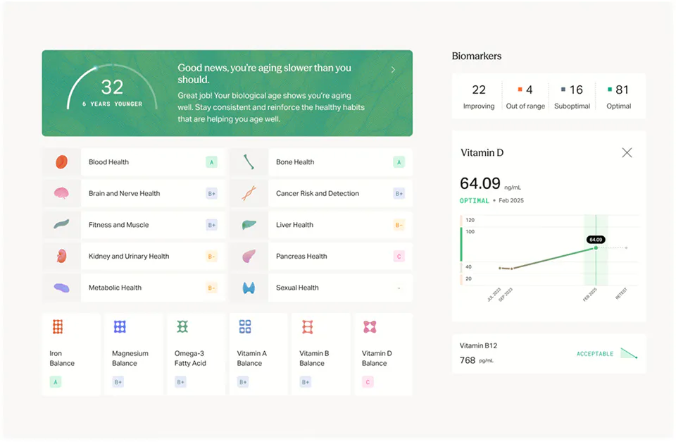
Insights that connect the dots. A clinician reads your whole picture, not one result in isolation, connecting family history, past results, and lifestyle into clear next steps.
Your personalized action plan. Nutrition, supplements, and training in one clinician-reviewed protocol, updated as your results change.
Track your trends. Upload results from any lab and see each biomarker trend over time, so you know what is working.
Never miss a recheck. Mito tells you what is worth retesting and when, then reminds you so nothing slips.
What to expect
- 1 Book instantly
Click, book, done. Choose a convenient lab location near you. Transparent, up-front pricing.
- 2 Quick lab visit
Testing to fit your busy schedule, usually 15 minutes or less. Walk-in and appointments available.
- 3 Fast, dashboard-delivered results
Your results post straight to your dashboard as soon as the lab completes them.
- 4 Expert guidance
Included with Mito membership. A clinician reviews your results and your personalized action plan follows, with clear next steps.
What members are saying
-
@nasdaily
-
15/10 experience - I felt actually taken care of
Super impressed with my Mito experience - the thoroughness of the intake, the data tracking and the doctor's visit.
RS Reena Sudan -
@ellarosemcfadin
-
Helped show an overall picture
I would highly recommend this service to anyone that wants a more in-depth picture of their health and labs. The platform is super user friendly.
BS Bo Sal -
@katelyn_kim
-
Specific and actionable advice
It felt like, for the first time, I had real data guiding me to make meaningful changes and I've definitely noticed the difference.
MH Maya Hickey -
@bryce_witmer
-
Tired of arguing with my PCP
After extensive research, I decided Mito was the best option. The insights were clear and make sense. Mito gave me peace of mind and a path to follow.
JO Josh -
@alexandradawson
-
3x the tests for less money
A great alternative to the basic blood work at your local clinic. Love the report card format. Much better value.
P Page -
@daniklass
-
Best $9 a month I spend
The action plan changes every time new results come in. I've actually never had a health company recommend doing less. Refreshing.
DP Devi Patel -
@natalieefer
-
Finally, prices that make sense
Every test is published. I can see what it costs and what I save as a member. No surprises.
KT Kenji Tanaka -
@tyler_sarkisian
Everything your health needs,
in one membership
-

Every test at our cost
Members pay our cost on every test, with lab fees passed straight through. The full receipt is itemized, never padded.
-

Continuous tracking, all in one place
Upload past labs and watch your trends over time. Every marker and visit lives in one longitudinal record, so all your care stays together.
-

Year-round clinician support
Text anytime and get clinician-reviewed answers. When you want to go deeper, 1:1 consultations are available at affordable rates.
All for $9/month
Order any test or consult without joining. For $9/month, members unlock member prices, trend tracking, and year-round clinician guidance.
Mito Membership
$9 /mo
cancel anytime
Without membership
$0
pay as you go
At-cost pricing on labs, scans, and more
Supplements at member rates NEW
On-demand clinician consults when you need guidance
Personalized action plans across supplements, exercise, nutrition, and sleep
AI health coaching to help you act on your results
Year-round medical support, with in-chat clinician escalation
All your health records in one personal vault, with trends and biological age tracking
Early access to new diagnostics and product releases
More tests to consider
Loading essentials…
Frequently asked questions
View all FAQsHow does pricing work?
Every test shows the member price next to the standard non-member price, so you can see what membership saves you. The member price is our cost, covering the lab and what it takes to run the service. It is never a profit on the test itself. Mito makes its money on the membership, not on marking up your tests. Membership is $9 per month or $99 per year, and any applicable lab order or draw fee is itemized before you pay.
Where do I get tested?
Choose an available partner lab at checkout. If your cart contains multiple blood tests, Mito helps consolidate the eligible tests at one lab when possible. At-home kits and scans follow the collection or appointment process shown on their product page.
Is this eligible for HSA/FSA?
Yes. Mito's lab tests are HSA/FSA eligible, so you can pay with pre-tax dollars.
Add the test to your cart and select "Pay with HSA/FSA" at checkout. You'll finish on the secure checkout page of Flex, our HSA/FSA payments partner, and Flex emails you an itemized receipt from notifications@withflex.com for your records.
Most Mito tests are auto-substantiated, so you usually won't need a Letter of Medical Necessity. If your HSA/FSA administrator asks for more, contact us and we'll help.
When will I get my results?
Your results appear in your Mito dashboard after the laboratory completes processing. A clinician then reviews them before your analysis and personalized action plan are finalized. Timing varies by test, and specialty assays and at-home kits may take longer.
Do I need a physician's order?
No. Mito provides the laboratory order when one is required, so you do not need to arrange a separate physician visit before purchasing.
How do availability and prices vary by state?
Available in 46 states and Washington, DC.
| States | Provider | Availability | Member price | Non-member price |
|---|---|---|---|---|
| All states and DC except HI, NJ, NY, and RI | Quest Diagnostics | Available | $7.15 | $10.01 |
| HI, NJ, NY, and RI | Not currently available |
Recently viewed
Loading essentials…